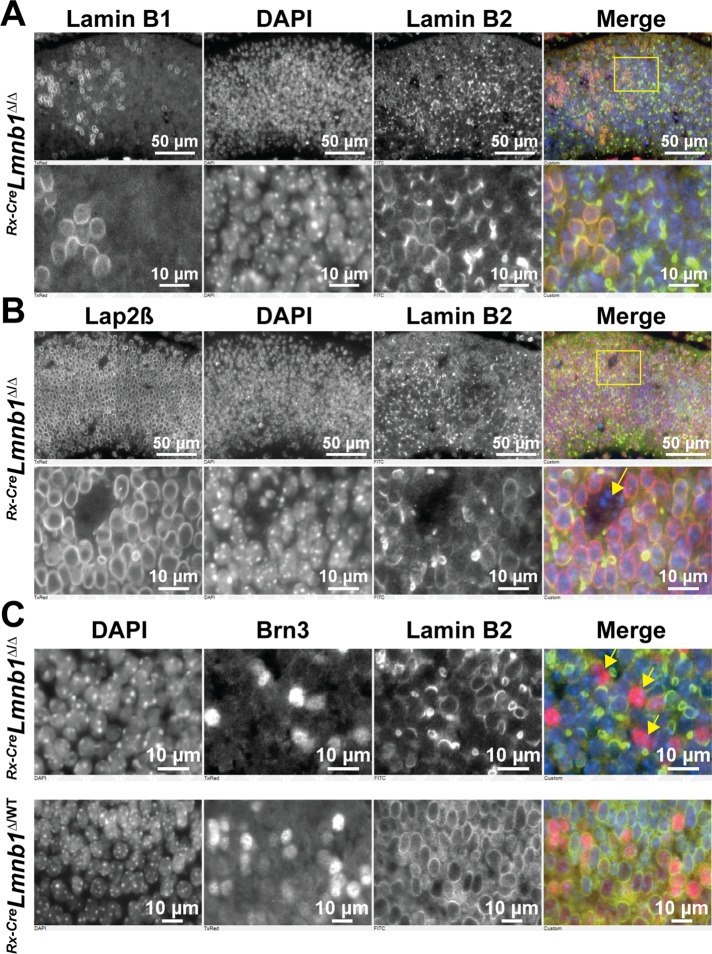
FIGURE 2:
Inactivation of Lmnb1 during embryonic development causes a collapse of the lamin B2 meshwork but does not prevent the birth of new postmitotic neurons. (A) Immunostaining of E14.5 Rx-CreLmnb1Δ/Δ retinas with lamin B1 and lamin B2. In the merged image, lamin B1 is red and lamin B2 is green; DNA was stained with DAPI (blue). Bottom, zoomed-in views of the yellow boxed area in top. (B) Immunostaining of E14.5 Rx-CreLmnb1Δ/Δ retinas with Lap2β and lamin B2. In the merged image, Lap2β is red and lamin B2 is green; DNA was stained with DAPI (blue). Bottom, zoomed-in views of the yellow boxed area in the top. The yellow arrow points to apoptotic nuclei in a gap. (C) Immunostaining of retinas from E14.5 Rx-CreLmnb1Δ/Δ (top) and Rx-CreLmnb1Δ/WT (bottom) mice with antibodies against lamin B2 and Brn3, a marker of RGCs. In the merged image, Brn3 is red and lamin B2 is green; DNA was stained with DAPI (blue). The arrows point to misshapen nuclei of RGCs. These studies show that RGCs devoid of lamin B1 are present and have irregularly shaped nuclei (yellow arrows). See also Supplemental Figure S2.

