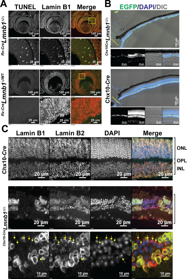
FIGURE 3:
Inactivation of Lmnb1 during embryonic development leads to increased apoptosis and reduced retinal cellularity in adult mice. (A) TUNEL labeling of E14.5 Rx-CreLmnb1Δ/Δ (top) and Rx-CreLmnb1Δ/WT (bottom) mice; specimens were also stained for lamin B1. Note the significant increase in apoptosis in retinal patches devoid of lamin B1. See also Supplemental Figure S3. (B) Low-power images of half-retinas from a Chx10-CreLmnb1Δ/Δ mouse (top) and a Chx10-Cre transgenic control mouse (bottom) stained with DAPI (blue). Cre recombinase expression in retinal bipolar cells is evident from EGFP expression (green). Bottom views emphasize regions of the retina that are devoid of photoreceptors. (C) Coimmunostaining for lamin B1 and lamin B2 of paraffin-embedded retinal sections from a Chx10-Cre transgenic mouse (top) and a Chx10-CreLmnb1Δ/Δ mouse (bottom). Note the asymmetric distribution of lamin B2 in a few cells with little or no lamin B1 (yellow arrows).

