FIGURE 4:
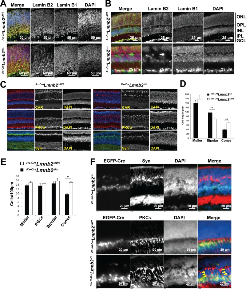
Lamin B2 is not required for neurogenesis but adversely affects lamination of retinal neurons. (A) Immunostaining of retinas from E14.5 Rx-CreLmnb2Δ/WT (top) and Rx-CreLmnb2Δ/Δ (bottom) mice with antibodies for lamin B1 and lamin B2. In the merged image, lamin B1 is red and lamin B2 is green; DNA was stained with DAPI (blue). Note the normal localization of lamin B1 in progenitors lacking lamin B2. (B) Immunostaining of retinas from P25 Rx-CreLmnb2Δ/WT (top) and Rx-CreLmnb2Δ/Δ (bottom) mice with antibodies against lamin B1 and lamin B2. Lamin B1 was normally positioned at the nuclear rim in all cells lacking lamin B2. In addition, in the DAPI-stained retina, note the intermixing of nuclei from the INL and ONL in patches of retina lacking lamin B2. IPL, inner plexiform layer; GCL, ganglion cell layer. (C) Immunostaining of retinas from P25 Rx-CreLmnb2Δ/WT (left) and Rx-CreLmnb2Δ/Δ (right) littermate mice with antibodies against CAR, PKCα (a marker of retinal bipolar cells), and synaptophysin (Syn). These images show abnormalities in the length of cone photoreceptors, length of bipolar cells, and ectopic synaptogenesis in retinas lacking lamin B2. (D) Length of Muller, bipolar, and cone photoreceptor cells in retinas of mice with the indicated genotypes. Measurements were performed in retinas of at least three mice per genotype. Data are represented as mean ± SEM. (E) Population counts of retinal cell types in retinas of P25 mice of indicated genotypes. Counts were performed on sections stained with cell type–specific markers (see Materials and Methods) using at least three retinas per genotype from at least two different litters. Data are represented as mean ± SEM. (F) Overview of frozen sections from Chx10-CreLmnb2Δ/Δ retinas immunostained with synaptophysin (top). Bottom, PKCα immunostaining of Chx10-CreLmnb2Δ/WT (top) and Chx10-CreLmnb2Δ/Δ (bottom) retinas, revealing abnormal elongation of apical axons from bipolar cells expressing EGFP-tagged Cre recombinase (arrowheads).

