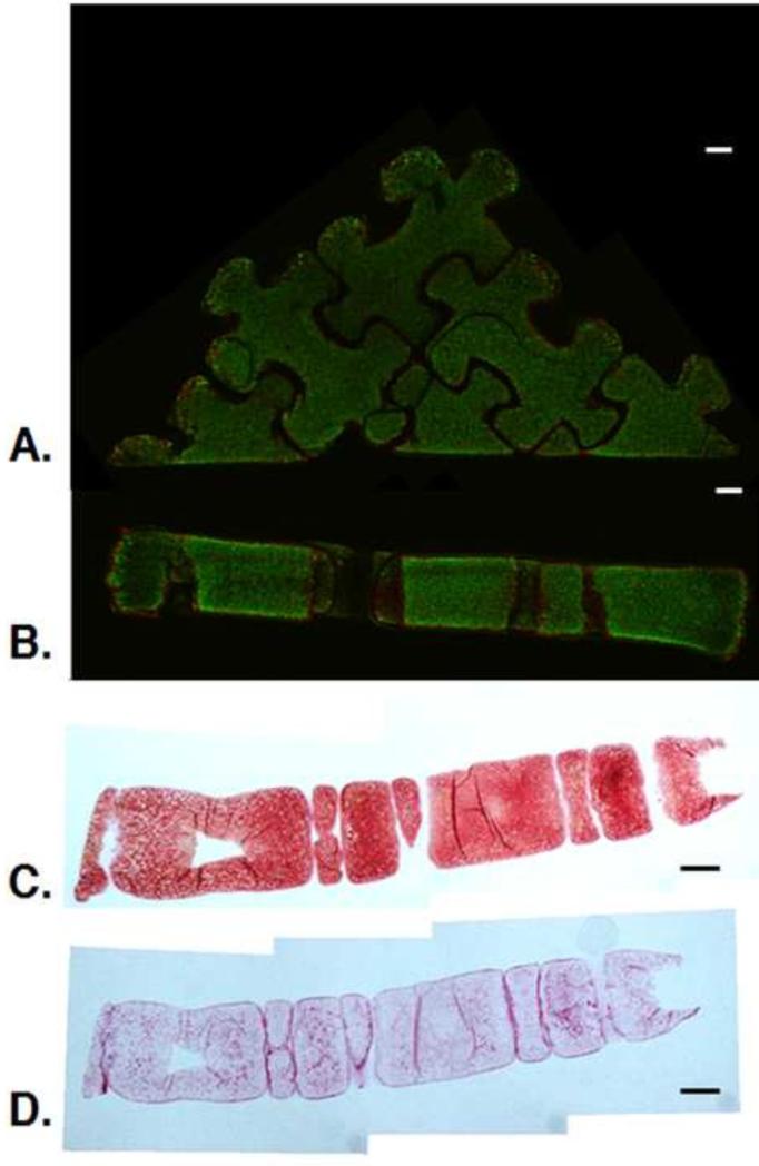
Fig. 6.
A-B. Live/Dead image of surface (A) and cross-section (B) of 3×3 puzzle arrangement at day 49. C-D. Cross-sectional histology of assembled 3×3 puzzle arrangement at day 49: Safranin O/Fast Green for GAG visualization (C) and Picrosirius Red for collagen visualization (D). (n=1). Scale bars: 1 mm. Live/Dead images provided to clarify orientation of histological sections.

