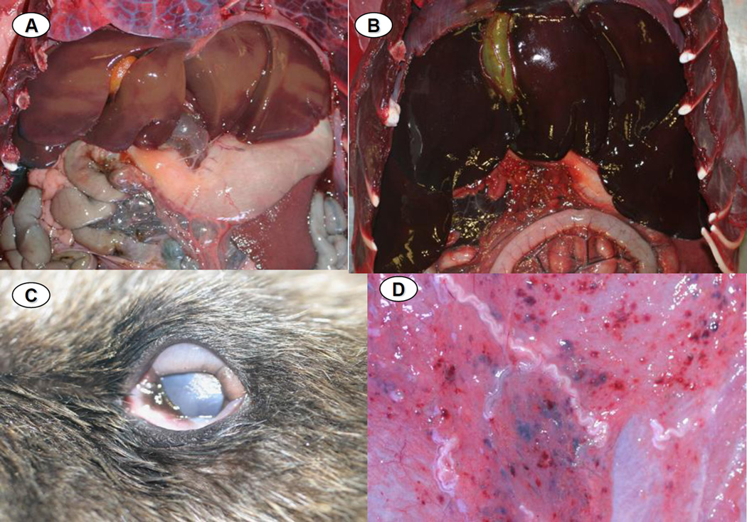
Figure 3.
A: Normal (control) sea otter liver, characterized by diffuse, pale tan-brown color and sharp lobular margins. 3B: Liver from an otter that died during the epizootic, showing severe, diffuse passive vascular congestion and rounded lobular margins, presumably due to cardiac insufficiency as a result of the myocardial lesions depicted in Figure 2 above. 3C: Diffuse chemosis affecting the ocular conjunctiva of an otter that stranded during the epizootic. 3D: Multifocal petechiation and mild serous atrophy of adipose of the ventral abdominal subcutis of an otter that died during the epizootic.

