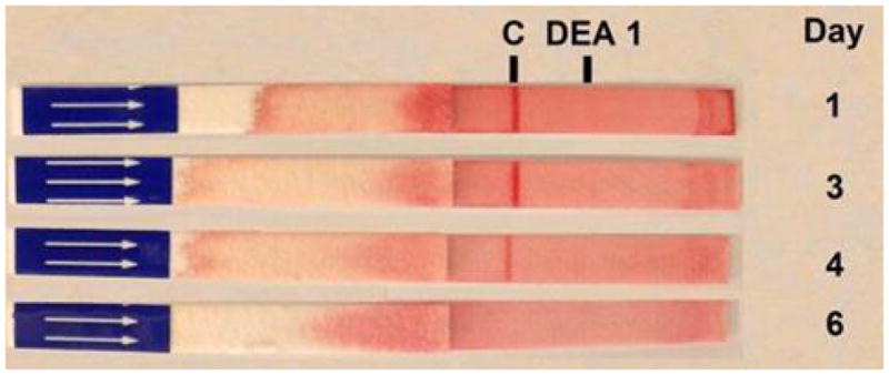
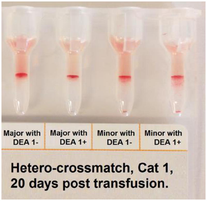
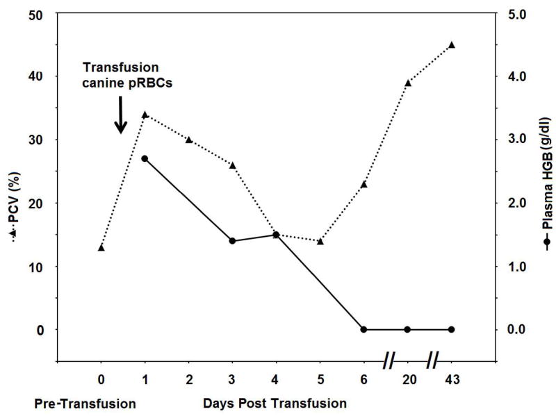
Figure 1.


Illustrative results from Cat 1, which received a xenotransfusion with canine pRBC (A). PCV and plasma HGB values (On day 0 the plasma appeared non-hemolyzed). (B) Microcapillary tubes showing PCV decline and hemolyzed plasma. (C) DEA 1 Strip showing a fading positive control band. (D) Crossmatch with canine blood, showing incompatibility for the major and minor crossmatch with DEA 1+ and - blood

