Figure 2.
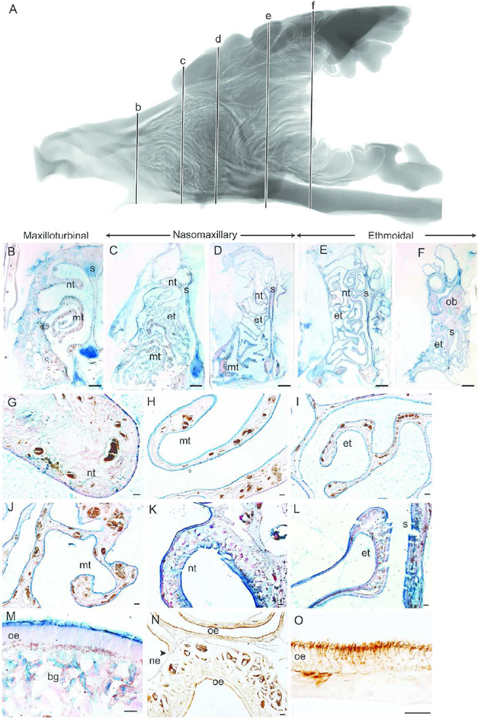
Epithelial lining of the bobcat nasal fossa. Sagittal view of segmented MRI scans of the bobcat nasal airway showing five coronal sections (b–f) selected for morphological and histological representation (A). The five coronal sections along the rostrocaudal axis illustrate the maxilloturbinal (B–C), nasomaxillary (D) and ethmoturbinal (E–F) regions of the bobcat nasal fossa. Magnified views show the nasoturbinal (G), maxilloturbinal (H, J) and ethmoturbinal (I) covered with nonsensory epithelium and packed with Alcian blue labeled goblet cells at rostral regions of the nasal fossa. Olfactory epithelium covered the nasoturbinal (K), septum (L), and ethmoturbinal (L) in more caudal regions and had the characteristic epithelial thickness and Bowman’s glands in the lamina propria (M) and labeled with β-tubulin III antibody (N–O). nt = nasoturbinal; mt = maxilloturbinal; s = septum; et = ethmoturbinals; ob = olfactory bulb; oe = olfactory epithelium; bg = Bowman’s gland; ne = nonsensory epithelium. Scale bar: B–F = 2 mm; G–L, N = 100 µm; and M, O = 50 µm.

