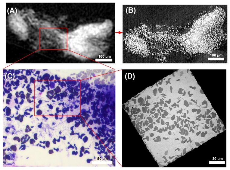
Figure 3.
Imaging sequence of a specimen which was determined to be invalid for the clinical study. (A) A slice through the bench-top micro-CT data shows an unorganized grainy texture. (B) The same slice from the higher resolution synchrotron CT data reveals that the unorganized grainy texture is discrete dots of osmium concentration. (C) The sample was sectioned to the location of the slice, and light microscopy image resolved the dots as red blood cells. (D) Transmission electron microscopy image of a section at the same location confirms the finding. The example illustrates how the bench-top scanner at 10 μm resolution provided sufficient information to exclude invalid samples. The higher resolution synchrotron CT helped to better understand the images from the bench-top scanners. The LM and TEM sections were necessarily separated by a few microns and the details of the LM and TEM images differ at the level of individual red blood cells.

