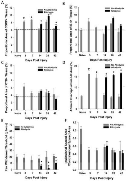
Figure 7. Quantitative analysis of primary afferents in the C7-8 dorsal horn.
Proportional area within the ipsilesional C7-8 dorsal horn was determined. Proportional area of positively labeled afferents within the dorsal horn revealed significant increases in CGRP (A) and IB-4 (B) but not CTB (C) immunoreactivity within the dorsal horn at 3 and 7 days post SCI (#p<.05 vs. naïve), as well as in the cohort of SCI rats that exhibit allodynia at 14, 28 and 42 dpi (*p<.05 vs naïve, SCI No allodynia at 14 dpi, dashed line indicates naïve levels). The area of nociceptive afferent overlap was significantly greater at 3 and 7dpi, and in SCI rats that have SCI-induced allodynia at 14, 28 and 42 dpi. A subset of SCI rats exhibited tactile allodynia at 14, 28 and 42 dpi (E, black bars, p<.05 vs naïve, 3 and 7 dpi, and No Allodynia 14, 28 and 42 dpi) that correlated with the increase in proportional area of CGRP+ and IB-4+ nociceptive afferent fibers in the dorsal horn. The increased immunoreactivity of nociceptive primary afferents was not associated with changes in the amount of tissue sparing at the lesion epicenter (F).

