
Figure 3. Kir2.1 channels are the major Kir channel isoform in ECs .
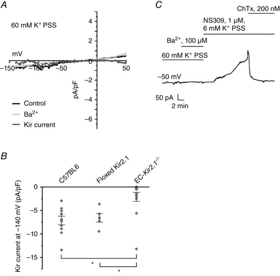
Kir currents were recorded using the perforated patch configuration of the patch‐clamp technique in ECs from third‐order mesenteric arteries. A, representative current traces in an EC from an EC‐Kir2.1 −/− mouse. The Kir currents were calculated as the difference between the traces before and after addition of Ba2+. B, a scatter plot compares the current densities at −140 mV between the ECs from the C57BL6 mice, floxed Kir2.1 mice, and EC‐Kir2.1 −/− mice (n = 5–12 ECs; *P < 0.001 using One‐Way ANOVA). C, NS309 was used to evoke outward IK/SK currents to confirm that the cell being studied was an EC.