
Figure 4. Combined stretch and electrical pacing augments HO‐1 but not nuclear MLP .
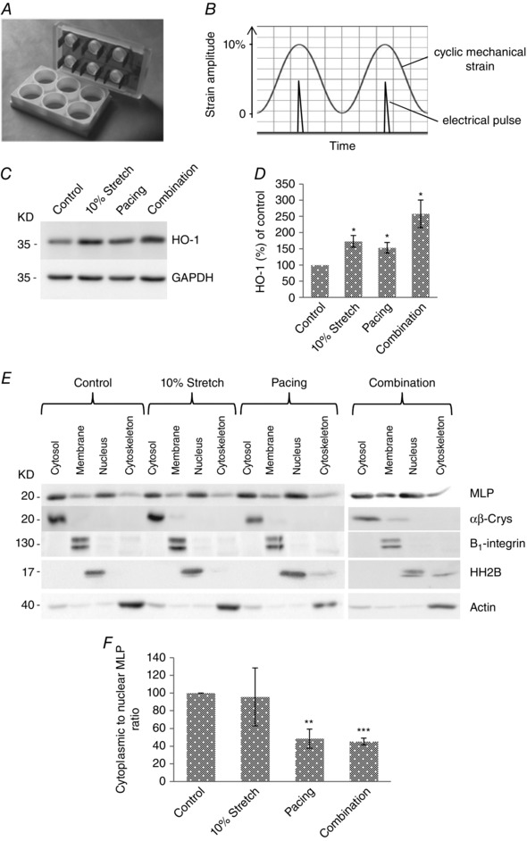
A, electrodes from the IonOptix C‐pace were inserted into the Flexcell six well dishes, allowing myocytes to be simultaneously stretched and paced. B, cells were stretched up to 10% maximal strain and the electrical pulse was applied to coincide with the point of maximal stain. C, western blot of HO‐1 and GAPDH in cultured neonatal myocytes following stretch, pacing and combined stimuli. D, quantification of HO‐1 protein following treatments. E, western blot to assess the subcellular distribution of MLP in cells following 10% stretch/strain, electrical pacing and combination of 10% stretch/strain and electrical pacing. αβ‐Crys, integrin β1, histine H2B and actin have been used as the marker of the cytoplasmic, membrane, nuclear and cytoskeletal pool of protein, respectively, to verify the fractionation process. F, quantification of MLP presented as cytoplasmic to nuclear ratio in cells following treatments. N ≥ 4 cultures.*P < 0.05, **P < 0.01 and ***P < 0.001 compared to control.