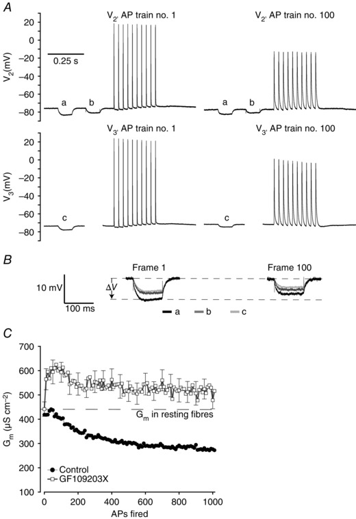
Figure 2. Three‐electrode determination of Gm and recordings of APs in PKC‐inhibited human abdominal muscle fibres .

A, representative recordings of the first (AP 1–10) and 100th (AP 991–1000) AP trains in a human abdominal muscle fibre in the presence of 1 μm PKC inhibitor (GF109203X). B, enlargements of the voltage deflections from the two runs of the current protocols shown in A that were used to determine cable parameters. C, average G m in human abdominal muscle fibres exposed to 1 μm GF109203X during excitation of 1000 APs as illustrated in A (n = 16). The comparable G m measurements from fibres without PKC inhibitor (Fig. 1 E) have been included to indicate the difference between the two experimental conditions. Average data are presented as means ± SEM.
