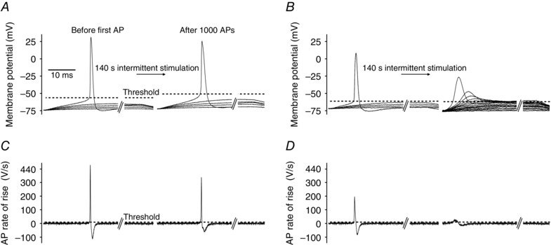
Figure 3. Illustration of AP threshold determination in human abdominal muscle fibres .

Rheobase current was determined before and immediately after firing 1000 APs (using the protocols illustrated in Fig. 1 B) by injecting 50 ms‐long current pulses that rose progressively from 10 nA in 5 nA steps until an AP was elicited. A, membrane potential recordings from a representative abdominal fibre during rheobase current measurement before and after firing 1000 APs (every second current trace was omitted for clarity). Dashed lines illustrate AP threshold identified as the voltage at which the AP rate of rise exceeded 10 V s−1 (Novak et al. 2015). B, as A but in the presence of 1 μm GF109203X. C and D, first derivative (dV/dt) of the membrane voltage in A and B in which the APs were triggered. The time point when the membrane potential reached AP threshold was identified as the first time point when dV/dt exceeded 10 V s−1 (dashed line).
