
Figure 3. Localization of FFA3‐IR in cholinergic nerves in whole mounts of mucosa and in submucosal plexus .
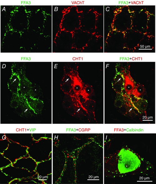
A–C, FFA3 (A) and VAChT (B) in mucosal plexus. Both FFA3‐ and VAChT‐IR nerves (merged image in C) surrounded the crypts. D–F, FFA3 (D) and CHT1 (E) expression in the submucosal plexus. Most FFA3‐IR nerve fibres and endings expressed CHT1 (arrows in F), whereas FFA3‐IR was faint in CHT1‐IR neuron somata (asterisks). G, CHT1 (red) and VIP (green) expressed in mucosal plexus. VIP‐IR and CHT1‐IR were detected in individual nerve fibres. H and I, FFA3 and α‐CGRP (H) or calbindin (I) in submucosal plexus. Calbindin‐IR neuron (asterisk) had no FFA3‐IR.