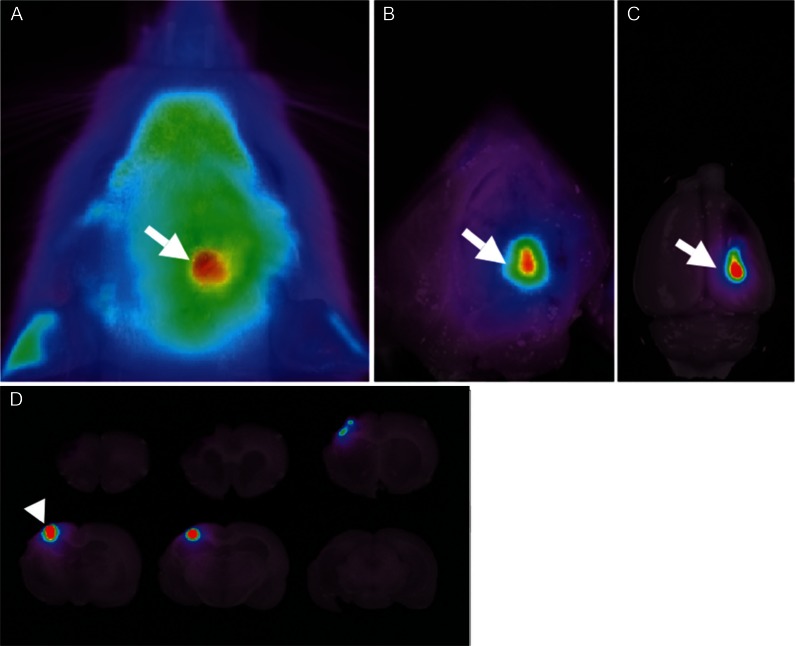
Fig. 4.
A: Near-infrared fluorescence bioimaging enables us to directly visualize the QD-labeled BMSCs through the scalp in the living rats subjected to permanent middle cerebral artery occlusion model for up to 8 weeks after direct transplantation. B–D: Post-mortem study reveals that the fluorescence emitted from the engrafted BMSCs can be observed through the skull (B) and cortical surface (C) and on coronal slices (D).45) BMSC: bone marrow stromal cell.

