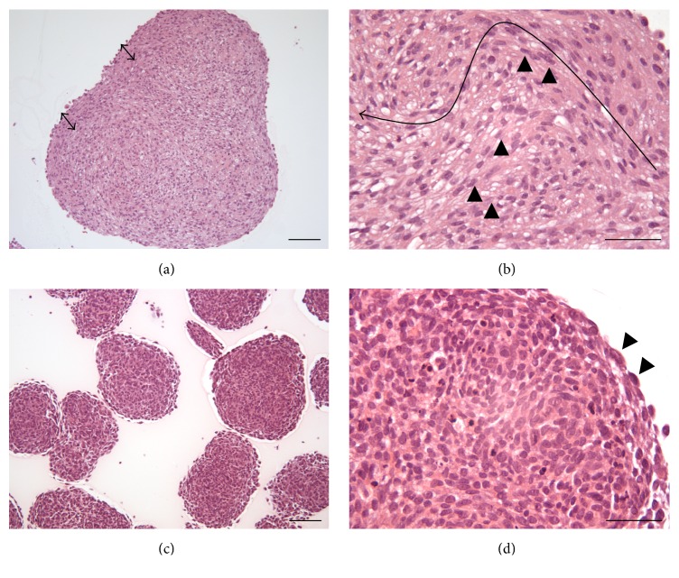
Figure 1.
Histological H&E stained sections of U-87 MG ((a), (b)) and LN-229 ((c), (d)) glioma cell spheroids grown for 14 days. (a) The arrows indicate the presence of a capsule-like structure in the periphery of the U-87 MG spheroids. (b) Cells in the periphery were orientated partially in a parallel manner and featured elongated spindle-shaped cells with longish nuclei that occasionally grew into the core of the spheroids (black arrow heads) and formed connective tissue like structures. The elongated arrow indicates the route from the periphery to the core. (c) Spheroids grown from LN-229 cells were less dense than U-87 MG spheroids. (d) The spheroid structure was homogenous and only a thin layer of flat cells was observed circumscribing the spheroid (black arrow heads). Scale bars: (a) and (c) 100 μm, (b) and (d) 50 μm.

