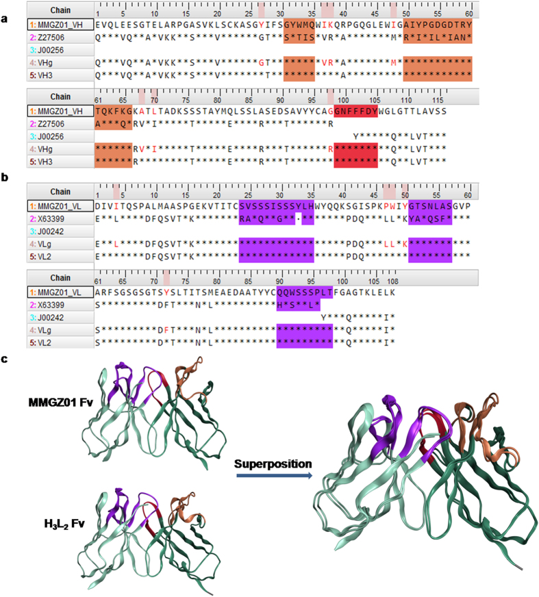
Figure 2. Amino acid sequences alignment and structure comparison between murine and humanized antibody.
(a) VH sequence alignment and (b) VL sequence alignment. MMGZ01_VH and MMGZ01_VL were named for VH and VL regions of MMGZ01 murine antibody. Z27506 and J00256 found in V and J genes were chosen as human FRs donors for the humanized VH. Similarly, X63399 and J00242 were chosen as human FRs donors for the humanized VL. VHg and VLg were VH and VL regions of CDR grafted antibody. VH3 and VL2 were VH and VL regions of the final version of humanized antibody. The residues colored red were back mutations in the final version. * represents residues that are identical to the corresponding residues of MMGZ01. CDR1-2 regions of VH are marked orange and CDR3 region of VH are marked red. CDR1-3 regions of VL are marked purple. CDR regions were identified with the Kabat numbering scheme17. (c) Superposed structure of MMGZ01 Fv and H3L2 Fv. The calculated structural RMSD was 0.824 Å.

