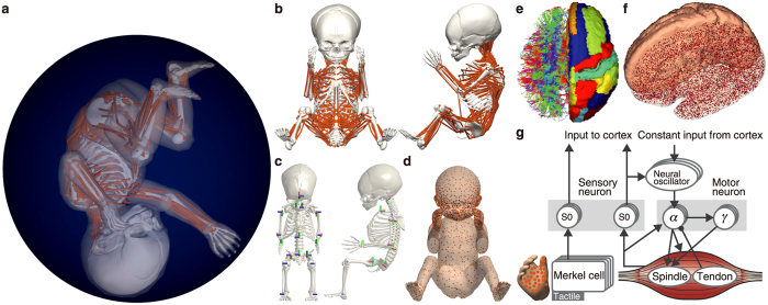
Figure 1. Overview of the embodied brain model of a human foetus.
(a) Foetal body model in the intrauterine condition. (b) Foetal musculoskeletal model. Each line is a uniaxial muscle actuator in the body. (c) Joint positions and directions. The cylinders represent the joint axes. (d) Tactile sensors throughout the body. The distribution was based on human two-point discrimination data. (e) Representative tractography and parcellation of preterm infant brain MRI scans. The colours on the left indicate the spatial relationships between the fibre end points. Red: left-right; blue: dorsal-ventral; green: anterior-posterior orientations. The colours on the right represent the brain regions defined in the brain atlas. (f) Model neurons in the cortical model. The dots represent neurons. (g) Spinal circuit model. The arrows and filled circles represent the excitatory and inhibitory connections, respectively. S0 represents sensory interneurons.

