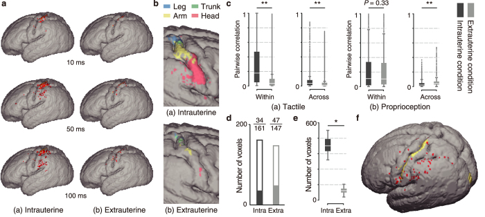
Figure 4. Cortical learning under intrauterine and extrauterine conditions.
(a) An example of the evolution of the cortical activities over time upon somatosensory input to the right arm. The coloured cortical regions represent the regions that significantly respond to the sensory input. (b) Cortical regions that significantly respond to specific single body parts. (c) Pair-wise correlation of the sensory feedbacks within and across body parts during simulated whole-body movements (**P < 10−24). (d) Numbers of voxels that significantly respond to proprioception feedback during whole-body movements. The white and coloured bars indicate voxels that respond to proprioception feedback from specific single body parts and across multiple body parts, respectively. (e,f) Cortical regions that significantly respond to simultaneous visual-somatosensory inputs, but not separate inputs. (e) The number of these cortical regions significantly increased in the learned cortical model inside the uterus (*P < 10−3). (f) The red regions are the cortical regions that responded to simultaneous inputs in the model learned inside the uterus. The yellow regions represent the cortical regions that received the sensory input.

