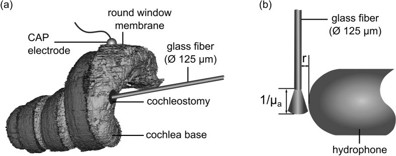
Figure 5.
(a) Segmented cochlea of a SLOT (scanning laser optical tomography) dataset with a cochleostomy inside the basal turn. An optical glass fiber was placed inside scala tympani and a CAP electrode was located at the round window membrane (RWM) for recording auditory responses as schematically shown. (b) Schematic illustration of the hydrophone and optical fiber position inside water as a sample medium for pressure measurements (not to scale).

