Figure 5.

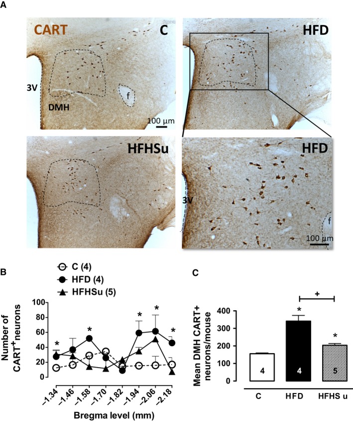
CART‐positive (CART+) neurones are higher in DMH of HFD mice. (A) Photomicrographs in light field at the DMH level of C (n = 4), HFD (n = 4), and HFHSu (n = 5) mice depicting the CART immunoreactive neuronal cell bodies. (B) Rostrocaudal distribution of CART+ in DMH from the bregma level among the groups. (C) Total number of CART+ neurones throughout the DMH. Fornix (f), third ventricle (3V). *P < 0.05 versus C, + P < 0.05 versus HFD; One‐way ANOVA with Newman–Keuls post hoc test.
