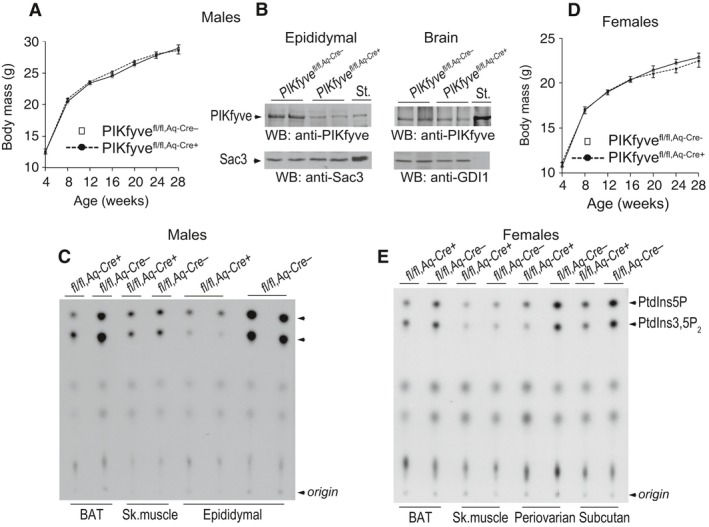
Figure 3.

Validation of fat‐specific ablation of PIKfyve in PIKfyvefl/fl,Aq‐Cre+ mice by Western blotting or in vitro kinase activity. (A and D) No differences in body weight gain of male (A, n = 12/group) or female PIKfyvefl/fl,Aq‐Cre+ mice (D, n = 7/group) fed a regular diet. (B) Clarified RIPA + lysates derived from fat (100 μg protein) or brain tissues (150 μg protein) dissected from male PIKfyvefl/fl,Aq‐Cre+ and PIKfyvefl/fl,Aq‐Cre− littermate mice, were examined by WB with anti‐PIKfyve, Sac3 or GDI1 (for equal loading) antibodies with a stripping step in between. PIKfyve was profoundly decreased in fat (by ~80%) but not in brain of PIKfyvefl/fl,Aq‐Cre+ compared to PIKfyvefl/fl,Aq‐Cre− controls. Shown are chemiluminescence detections from representative experiments with 2 mice/genotype for each tissue out of three to five independent determinations. St., PIKfyve or Sac3 standards from lysates of COS7 cells overexpressing these proteins. (C and E), Clarified fresh RIPA + lysates (600 μg protein), derived from the indicated tissues dissected from PIKfyvefl/fl,Aq‐Cre+ and PIKfyvefl/fl,Aq‐Cre− male (C) or female (E) mice underwent immunoprecipitation (IP) with anti‐PIKfyve antibodies for measuring the in vitro lipid kinase activity. Shown are representative autoradiograms of a TLC plate with resolved radiolabeled lipids demonstrating that in the fat depots of the PIKfyvefl/fl,Aq‐Cre+ mice, but not in the other tissues, synthesis of the PIKfyve lipid products were profoundly reduced.
