Figure 6.

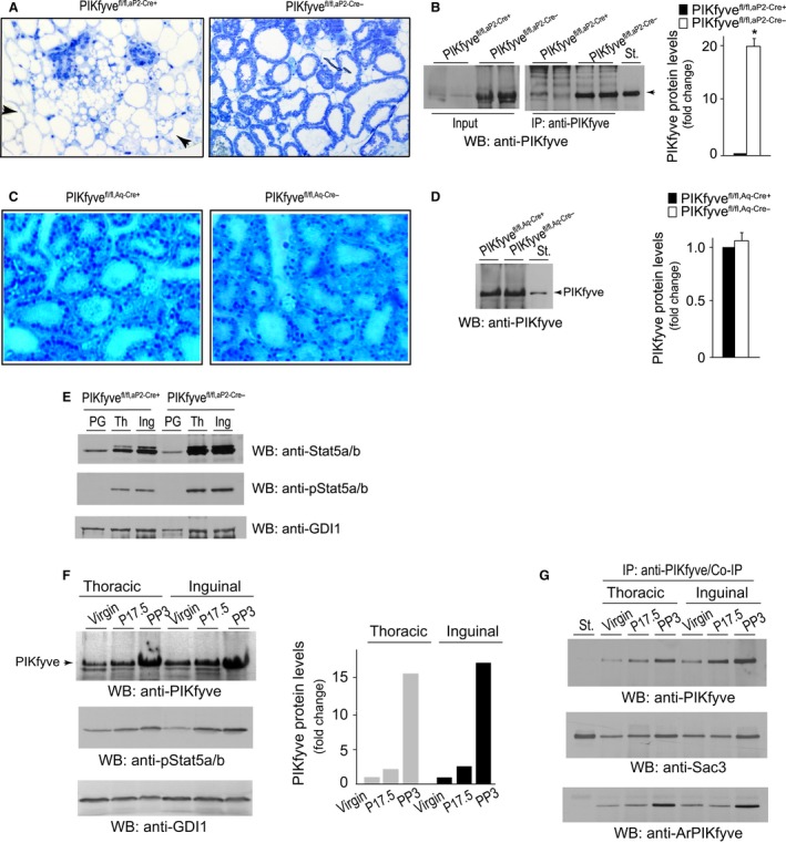
Pikfyve disruption by aP2‐Cre, but not by Aq‐Cre, causes severe mammary phenotype. (A) Histology of formalin‐fixed plastic‐embedded stained sections of mammary glands (#4, inguinal) derived from 9‐week‐old PP3 PIKfyvefl/fl, aP 2‐Cre+ and PIKfyvefl/fl, aP 2‐Cre− primiparous females. Apparent is severely underdeveloped gland with few lobuloalveolar structures within the bulk of mammary fat cells in PIKfyvefl/fl, aP 2‐Cre+ dams (arrows). Alveolar lobuli of control PIKfyvefl/fl, aP 2‐Cre− glands are well‐expanded and milk‐filled. (B) Clarified RIPA + lysates (150 μg protein) from mammary glands dissected from 9‐week old PP3 PIKfyvefl/fl, aP 2‐Cre+ and PIKfyvefl/fl, aP 2‐Cre− primiparas were examined by WB with anti‐PIKfyve antibodies directly (left panel) or subsequent to IP with anti‐PIKfyve antibodies (right panel). Shown are chemiluminescence detections from a representative experiment with two mice/genotype of three determinations. Graph, quantified changes in PIKfyve levels between the two genotypes. *P < 0.001, n = 3. (C) Histology of formalin‐fixed plastic‐embedded stained sections of mammary glands (#4, inguinal) derived from 9‐week‐old PP3 PIKfyvefl/fl,Aq‐Cre+ and PIKfyvefl/fl,Aq‐Cre− primiparas, showing similar lobuloalveolar structures in the two genotypes. (D) Clarified RIPA + lysates (150 μg protein), derived from mammary glands dissected from 9‐week‐old PP3 PIKfyvefl/fl,Aq‐Cre+ and PIKfyvefl/fl,Aq‐Cre− primiparous sisters were examined by WB with anti‐PIKfyve antibodies. Shown is a chemiluminescence detection from a representative experiment out of three with similar results and quantification. (E) Clarified RIPA 2+ lysates (40 μg protein) obtained from perigonadal fat (PG) or thoracic (Th) and inguinal (Ing) mammary glands dissected from 20‐week‐old PP3 PIKfyvefl/fl, aP 2‐Cre+ and PIKfyvefl/fl, aP 2‐Cre− primiparous sisters were examined by WB with anti‐Stat5 and anti‐phospho‐Stat5 antibodies with a stripping step in between. The blot was reprobed with anti‐GDI1 to normalize for loading. Shown are chemiluminescence detections from a representative experiment out of two with similar results. (F) RIPA 2+ lysates were obtained from three PIKfyvefl/fl littermate females at the stage of virgin, pregnancy day 17.5 (P17.5) and postpartum day 3 (PP3). Clarified lysates were analyzed by WB (150 μg protein) with the indicated antibodies (left panel). Shown are chemiluminescence detections from a representative experiment probed with anti‐PIKfyve (the upper half of the membrane) and anti‐phospho‐Stat5 antibodies (the lower half of the membrane). The lower half of the membrane was reprobed with anti‐GDI1 to normalize for loading. The increase in PIKfyve levels is quantified from two independent experiments with similar results. (G) Lysates (600 μg) obtained as in F were analyzed by immunoprecipitation with anti‐PIKfyve antibodies to quantify the interaction partners Sac3 and ArPIKfyve and their association with PIKfyve. Shown are chemiluminescence detections from a representative IP experiment probed with the indicated antibodies subsequent to stripping. St., in B, D, & G, PIKfyve, Sac3, or ArPIKfyve standards from lysates of COS7 cells overexpressing the proteins.
