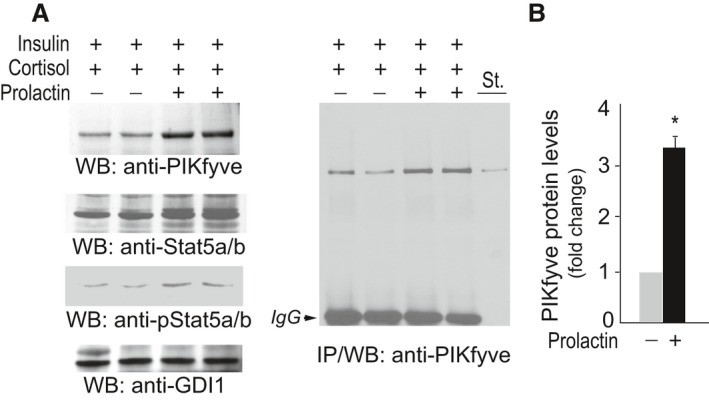
Figure 7.

PIKfyve is upregulated by PrlR activation in mammary gland explants of midpregnant mice. (A) Mammary gland explants derived from 6 to 8 midpregnant mice were cultured for 24 h with 1 μg/mL insulin and 10−7 mol/L cortisol. Incubation was then continued for additional 24 h after addition of Prl (1 μg/mL) where indicated. Clarified RIPA 2+ lysates from duplicate cultures were analyzed by WB (40 μg protein) with the indicated antibodies (left panel) and immunoprecipitation (IP) with the PIKfyve antibodies (right panel). St., lysates from COS7 cells overexpressing PIKfyve. Shown are chemiluminescence detections from representative WB and IP out of four independent experiments in duplicates. The membrane was reprobed with anti‐GDI1 to normalize for loading. (B) Quantification of the PIKfyve increases upon PrlR stimulation. *P < 0.001 versus prolactin nonstimulated mammary explants.
