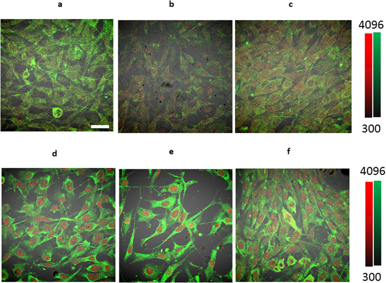
Figure 1. Simulation of tumor-associated pH environment in vitro.
Confocal imaging of rabbit polyclonal anti-OCT4 (red) staining and anti-vimentin (green) in 3T3 cells under various conditions. Fibroblasts (a) incubated in pH 7.4 media, (b) co-cultured with MDA-MB-231 cells in pH 7.4 media, (c) co-cultured with BXPC-3 in pH 7.4 media, (d) incubated in pH 6.5 media, (e) co-cultured with MDA-MB-231 cells in pH 6.5 media, (f) co-cultured with BXPC-3 breast cancer cells in pH 6.5 media. All the cells were incubated for 7 days with no changes in media. Magnification is 20x. Scale bar is 100  m.
m.

