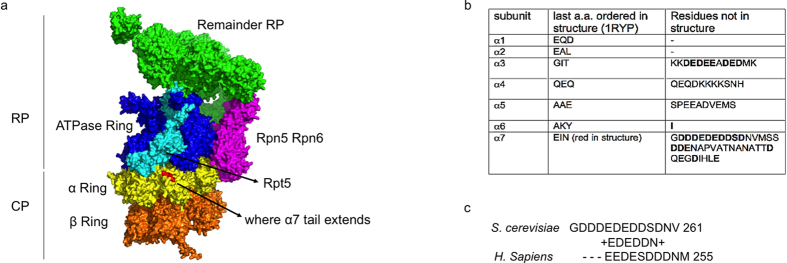
Figure 1. α7 as the putative binding site for Ecm29.
(a) CryoEM-based model of the Proteasome29 (PDB- 4B4T) showing regulatory particle (RP) and core particle (CP). Rpt5 is displayed in cyan and other Rpt subunits in blue. Lid subunits Rpn5 and Rpn6 that interact with the CP are shown in purple and the remainder of RP is shown in green. The core particle α subunits are colored in yellow, with the last three ordered amino acids of α7 shwon in red. β subunits are colored in orange. (b) Amino acids sequences from the α-ring subunits beyond crystal structure30 and model presented29. Second column shows the last three ordered amino acids for each α subunit. Third column shows C-terminal amino acids that were not resolved in the structure. Consecutive acidic residues in C-terminal of α7 residues that forms the acidic patch are underlined as red line. (c) Sequence alignment of acidic patch in the C-terminal tail of α7 from S. cerevisiae and H. sapiens.

