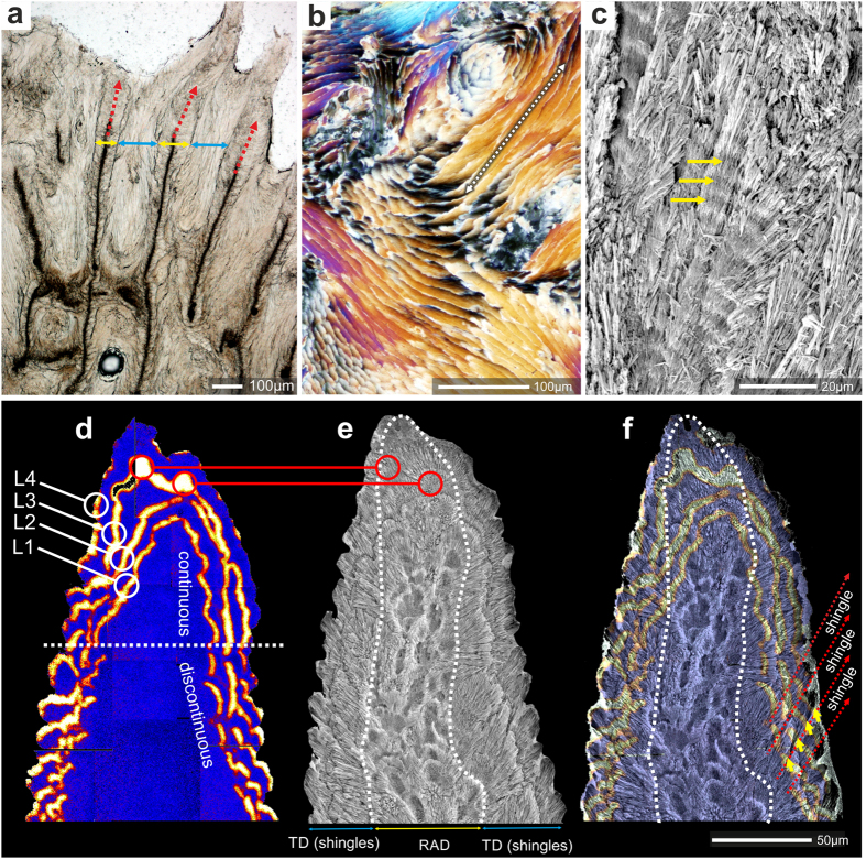
Figure 2. Microstructure and differentiation of shingles near the tip of coenosteal spinulae visualized by 86Sr labeling in Recent Acropora (A. eurystoma, ZPAL H.25/118(966)).
(a) longitudinal section along the fast growing skeletal regions includes spinulae (yellow arrows), RAD (red arrows) and shingles (blue arrows). Ultra-thin transverse section (b), polarized light)) and polished, slightly etched section (c) reveal longitudinal sections of shingles bundles of fibers several hundreds of micrometers long (dashed arrow in (b)), suggesting continuous growth of individual shingles, (d) NanoSIMS 86Sr/44Ca isotope mosaic map, (e) SEM image of polished and etched sample, (f) NanoSIMS and SEM images overlaid. 86Sr-labeling pulses (12 hours, separated by 36 hours, orange-yellow color) are continuous in the distal part of the spinula and discontinuous below, where shingles are forming. Blue regions represent skeleton with normal 86Sr/44Ca ratio. Red lines with circles in d an e indicate RAD labeled with 86Sr. Dashed white line in e and f outlines the RAD region.

