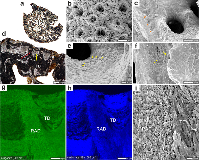
Figure 4. Example of good preservation of skeletal features (mineralogy, surface texture and microstructure) in fossil Acropora.
Acropora sp. ZPAL H.27/20(C21), Oligocene (Chattian), Saint-Paul-lès-Dax, France. (a) transverse section of corallum branch with white rectangle (enlargement in (d)) indicating region analyzed with micro-Raman (g,h); shingled thickening deposits marked with yellow and region of rapid accretion deposits with red arrows. Surface of the branch with lateral corallites and coenosteum (b). Enlargment of the calice surface (c) with desmocyte attachment scars (orange arrows). Shingled thickening deposits, (yellow arrows in (e,f)) are still discernible, although this feature usually is the first that is eroded. Micro-Raman maps (g,h) of region indicated in d: aragonite lattice mode at 203 cm−1 (g), and carbonate vibrational mode at 1085 cm−1 (h) to show that skeleton is entirely aragonite. RAD are composed of more compact skeletal tissue in comparison to TD deposits and consequently epoxy impregnated only TD deposits (black areas), (i) contact zone (polished and etched surface) between RAD and TD (shingles) deposits. Note regular increment lines in RAD and elongated bundles of fibers composing TD.

