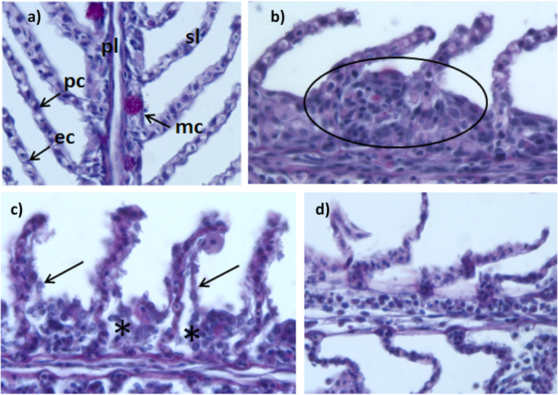
Figure 1. Gill lesions in fish exposed to Pelagia noctiluca.
(a) Control fish gills with unaltered primary lamellae (pl) with mucous cells (mc) and elongated secondary lamellae (sl) with flat epithelial cells (ep) and pillar cells (pc), (400×); (b–e). Pathological features in fish gills from the treatment groups after 8 h exposure to jellyfish (400×): (b) Hyperplasia of primary lamella; (c) Moderate lifting of epithelial cells (*) and cellular degeneration (arrows); (d) Absence of respiratory epithelium and loss of structure.

