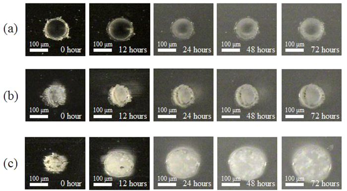
Figure 5. Digital microscopic images of the craters formed on PLGA films.
The time shown in each figure indicate the time of the samples immersed in PBS at 37 °C. Scale bars represent 100 μm. (a) The crater formed by mechanically milling. (b) The laser ablation crater under the condition of 800 nm, 1.0 J/cm2, 15000 pulses. (c) The laser ablation crater under the condition of 400 nm, 0.15 J/cm2, 15000 pulses.

