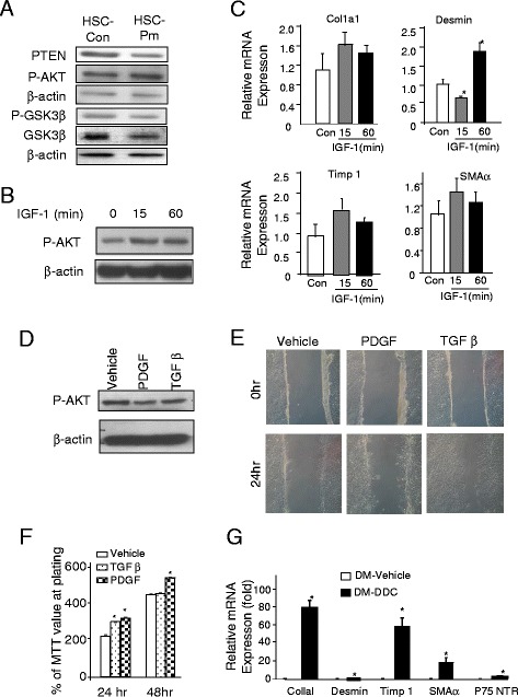
Fig. 6.

PTEN regulates HSC gene expression independent of AKT signal. a. Protein analysis of HSCs isolated from control (HSC-Con) and Pten null (HSC-Pten null) livers. Membrane is probed with PTEN, p-AKT, p-GSK3β, GSK3β, and α-actin for loading control. b Induction of AKT phosphorylation by IGF-1 treatment in human HSC cell line. β-actin is used as loading control. c Quantitative PCR analysis of expression of SMAα, Col1aI, Timp 1, and desmin in vehicle- and IGF-1 (gray bar, 15 min; and black bar, 1 h)-treated samples, respectively. n = 3, *p < 0.05 when compared to the vehicle-treated group. d Analysis of AKT phosphorylation in response to PDGF (50 ng/ml) and TGFβ (4 ng/ml) treatment. e PDGF and TGFβ treatment induced HSC migration as measured with a wound healing assay. Representative images of three experiments. f PDGF and TGFβ both induced cell proliferation in cultured HSCs. Cell proliferation is measured by a MTT assay. n = 3, *p < 0.05. g Relative expression of SMAα, Col1aI, Timp 1, p75NTR, and desmin in livers of Pten and Akt2 double mutant (Dm) mice treated with (black bar) or without DDC (open bar). n = 5, *p < 0.05 when compared to the vehicle-treated group
