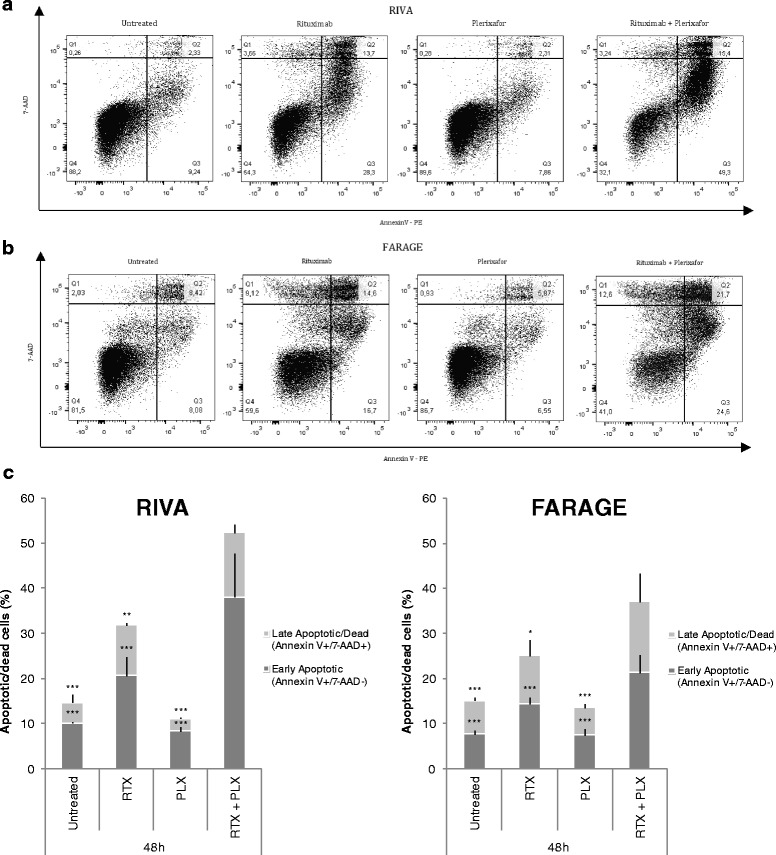
Fig. 3.

Addition of plerixafor to rituximab treatment significantly increased the fraction of apoptotic cells. The anti-CD20 antibody rituximab (10 μg/mL) and/or the CXCR4 antagonist plerixafor (500 μM) were administered to the diffuse large B-cell lymphoma (DLBCL) cell lines RIVA (a) and FARAGE (b). The cells were subjected to flow cytometry-based analysis 48 h later, using a combination of 7-AAD and PE-conjugated Annexin V. One representative experiment per cell line is shown, with numbers indicating the percentage of cells in each quadrant. (c) The fractions of early apoptotic and late apoptotic/dead cells are summarized as mean of two independent experiments ± SEM, with each experiment containing technical triplicates. Statistical significance between groups was determined using a two-level linear mixed effects model. *p < 0.05; **p < 0.01; ***p < 0.001; RTX, rituximab; PLX, plerixafor; Q1, dead cells (AnnexinV−/7-AAD+); Q2, late apoptotic/dead cells (AnnexinV+/7-AAD+); Q3, early apoptotic cells (AnnexinV+/7-AAD−); Q4, living cells (AnnexinV−/7-AAD−)
