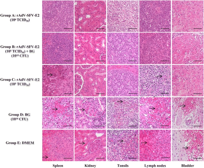
Figure 3.

Representative histopathological changes in pigs challenged with CSFV Shimen strain. Five groups of pigs (n = 4) were immunized and challenged as described in the “Materials and methods” section. At 15 days post-challenge (dpc), various tissues (spleen, kidney, tonsils, lymph nodes and bladder) were collected from the challenged animals, fixed with buffered 4% formalin and subsequently embedded in paraffin wax. Tissue sections (around 4-μm thick) were prepared and stained with hematoxylin and eosin for histopathological examinations. In Groups D and E, severe hemorrhages in the lymph nodes, tonsils, spleen, kidney and bladder were indicated as arrows; in Group C, slight to moderate histopathological changes were found in some tissues, such as focal necrosis in the splenic parenchyma (arrow) and depletion of lymphocytes (arrow) in the white pulp in the spleen and hemorrhages (arrow) in the lymph nodes. All the pigs in Group A or B did not show any histopathological changes. CFU: colony forming units. Bars 50 μm.
