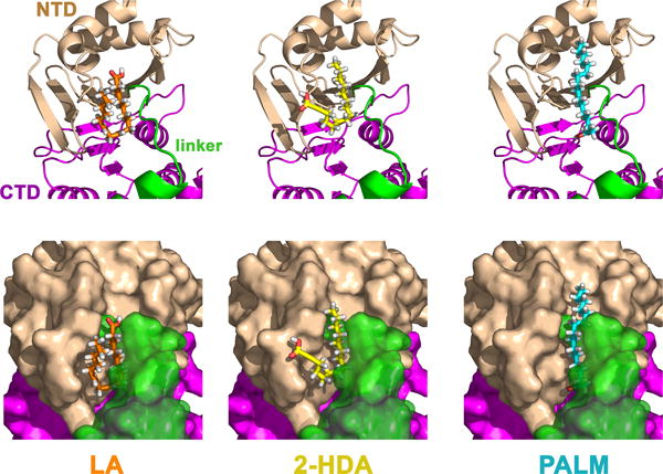
Figure 6. Blind docking of fatty acids into the molecular model of TrwD.

Blind docking predictions between a molecular model of TrwD (Ripoll-Rozada et al., 2012) and fatty acid ligands (linoleic acid (LA), 2-hexadecanoic acid (2-HDA), and palmitic acid (PALM)), were performed using the EADock dihedral spacing sampling engine of the Swiss-dock server (Grosdidier et al., 2011). Most of the binding poses clustered at a pocket localized at the interface between the N-terminal domain (NTD, wheat) and the linker region (green), which connects the NTD with the catalytic C-terminal domain (CTD, purple). The binding modes with the best energy and Full-Fitness are shown. Upper and bottom panels correspond to the same views in cartoon and surface representations, respectively. For clarity, transparency was applied to the linker region (green) in the surface representations (bottom panel).
