Fig. 5.
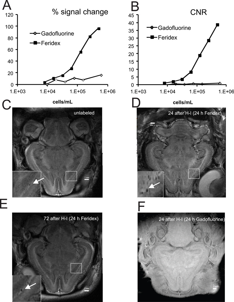
Cells were labeled with T2-shortening superparamagnetic iron oxide MRI contrast (Feridex) or T1- shortening contrast Gadofluorine M. Signal change (A) and contrast-to-noise ratio (B) determined on T2-weighted images for Feridex and T1-weighted images for Gadofluorine on agarose gel phantom with serial cell dilutions on 4.7T.
Detection of intravenously infused HUCBC on MRI in vivo. Rabbit kits were imaged on 4.7T magnet 24 hours after infusion of media (C, T2-weighted) or Feridex labeled cells (D, T2-weighted) 24 hours or 72 hours (E) after E29 H-I. F.- T1-weighted image of kit infused with Gadofluorine labeled cells 24 hours after E29 H-I. 2.5x10⁶ HUCBC cells were delivered by a PHD 2000 programmable pump. (Harvard Apparatus Holliston, MA).

