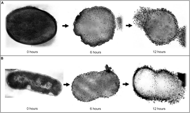
FIGURE 4.
Transmission electron microscope (TEM) images which visualize the morphological changes in bacteria upon treating with Kan-AuNPs at different intervals of time. (A) Represents sequential images (from left to right) of Gram-positive, S. epidermidis bacteria treated with Kan-AuNPs (18.00 μg mL-1) after 0, 6, and 12 h of incubation. (B) Represents sequential images (from left to right) of Gram-negative, E. aerogenes bacteria treated with Kan-AuNPs (16.00 μg mL-1) after 0, 6, and 12 h of incubation. After 6 h of exposure, Kan-AuNPs were found to adhere and penetrate the bacterial cell wall which resulted in disruption of cellular environment leading to lysis of cell due to leakage of cellular components as observed after 12 h of exposure. The results were similar for both Gram-positive and Gram-negative bacteria.

