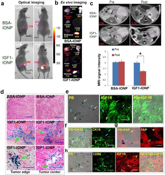
Figure 4.
Detection of targeted delivery of IGF1-IONPs into orthotopic pancreatic PDX tumors by optical imaging, MRI, and Prussian blue staining. (a) NIR optical imaging of whole body 24 h after IONP administration. Optical images were overlaid with X-ray images of the mice. Red numbers shown are the mean signal intensities of tumor areas. (b) Ex vivo optical imaging of representative tumors and organs following sacrificing the mice. Optical images were overlaid with bright-ffeld images to show location and size of tumors and organs. Numbers shown were the mean optical signal intensities of tumors or organs. (c) Pre and post 24 h T2-weighted MR images. Numbers shown are relative mean MRI signal intensities of the entire tumor. Bar figure shows quantification of MRI signals in the tumors prior to and 24 h after administration of difierent IONPs. Relative MRI signal was defined as the mean intensity of the tumor divided by the mean intensity of the muscle on the same MR image. *p < 0.0001. Pink arrows indicate the location of pancreatic PDX-tumor lesions. (d) Prussian blue staining of frozen tumor sections indicating the presence of IONPs in both tumor edge and tumor center (green arrows) after IGF1-IONP administration via the tail vein. IONPs were not detectable in the tumor treated with nontargeted BSA-IONPs. Blue: IONP-positive cells. Red: Nuclear fast red. (e) IGF1R antibody labeled tumor tissue sections obtained from IGF1-IONP-treated mice dual stained with Prussian blue staining. IONPs (blue) were detected in IGF1R highly expressing tumor cells (white arrows) and intermediate IGF1R expressing tumor stromal cells (light blue arrows). (f) Blue IONP-positive cells detected in CK19-positive ductal tumor cells (white arrows) and CK19 negative tumor stromal cells (light blue arrows). (g) Prussian blue staining positive cells found in FAP-positive stromal fibroblasts (light blue arrows). Yellow arrows indicate tumor cell areas. (h) IGF1-IONP-treated tumor tissue section doubly labeled with IGF1R (red) and CD68 (green) and then stained with Prussian blue (blue). IONP-positive cells were detected in CD68-positive macrophages (white arrows).

