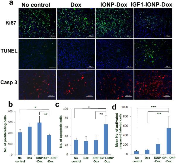
Figure 7.
Evaluation of the effects of IGF1-IONP-Dox treatment on cell proliferation and induction of apoptotic cell death in the pancreatic PDX tumors. (a) Immunofluorescence labeling of tumor tissue sections for cell proliferation marker (Ki67, green), TUNEL assay (green), and apoptotic cell death (active caspase-3, red). Hoechst 33342 nuclear stain (blue) was used to identify total cell populations in tumor tissue sections. (b-d) Quantitative analysis for (b) Ki-67-positive cells, (c) apoptotic cells, and (d) cleaved active caspase-3-positive cells from six randomly selected microscopic ffelds of tumor sections by ImageJ. *p < 0.03; **p < 0.007; ***p < 0.0001. Scale bars are 100 μm.

