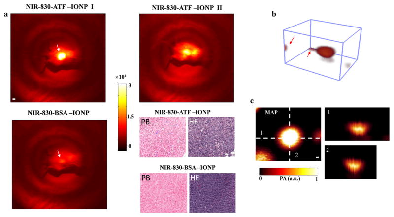
FIG. 2.
Validation of targeting ability and contrast enhancement for PAI of NIR-830–ATF–IONPs. a NIR fluorescence imaging of mice that received an injection of NIR-830–ATF–IONPs or NIR-830–BSA–IONPs. All tumors from the targeted animal group had strong optical signals and some showed strong signals from the internal organs (kidney and liver), possibly due to the non-specific uptake of the IONP by the RES in the liver and elimination of degraded NIR-830-dye-conjugates in the kidney. PB-stained tumor tissue sections from the mice injected with NIR-830–ATF–IONPs showed a cluster of blue-stained IONPs, while no blue-stained IONPs were found in the mice injected with NIR-830–BSA–IONPs. b A typical three-dimensional photoacoustic image with two tiny markers in the corners. Markers indicated by the red arrows were used to assistant surgeons in locating the tumors in the imaging area. c MAP and cross-sections of photoacoustic image shown in b. The MAP images were used to depict the surgical profile in the imaging area, and cross-sections provided depth information and axial dimension of tumors. NIR near-infrared, IONPs iron oxide nanoparticles, RES reticuloendothelial system, MAP maximum amplitude projection

