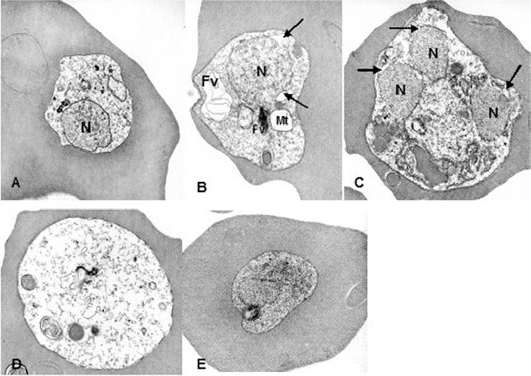
Figure 2.
Electron micrographs of P. falciparum (ItG2) parasites cultured in Apo E4/4 (A~D). Parasites infected with RBC exhibited mitochondrial swelling and dilatation of the perinuclear space. Cytoplasm became electron lucent, and membranous structures accumulated in the food vacuole. Micrograph E shows a control parasite cultured in Apo E 3/3. N; nucleus, FV; food vacuole, Mt; mitochondrion. Arrows indicate dilated perinuclear spaces.

