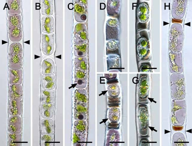
Figure 2.
Light micrographs of vegetative cells (A–C, F–H) and aplanospores (D, E) of Zygogonium SCOT (A, C–H: Zygogonium SCOT-p, B: Zygogonium SCOT-g). (A), (B) H-shaped cell wall structures (arrowheads). (C) One spherical dark purple inclusion per cell (arrow), dentate longitudinal cell walls. (D) Small aplanospores; brownish or purple granular cytoplasmic residue outside the spores. (E) Aplanospores embedded in brownish cytoplasmic residue and; numerous transparent particles in the periphery of the spore (arrows). (F) Encysted cells with two prominent chloroplasts. (G) Encysted cells surrounded by ‘highly ordered’ spherical bodies (arrows). (H) Purple filament formed by germination of aplanospores as indicated by the brownish remnant of cytoplasmic residue; H-shaped cell wall structures (arrowheads). Bars = 20 μm.

