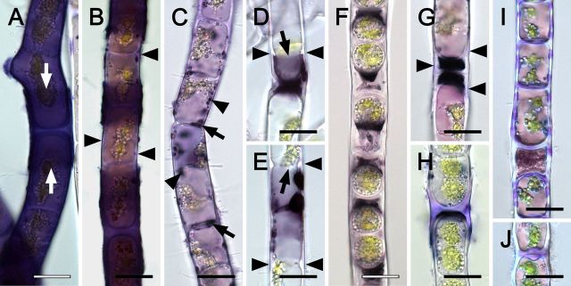
Figure 3.
Haematoxylin staining of Zygogonium AUT-p (A–G) and Zygogonium AUT-g (H–J) filaments. (A) Strong violet staining in the cell and in H-shaped cell wall structure (arrows). (B) Lilac (arrowheads) and dark violet to blackish staining in the cell wall. (C) Dark violet to blackish staining inside the protoplast (arrowheads) and cross cell walls (arrows). (D, E) Strong and sharply delineated staining in the protoplast near cell poles (arrow); deposition of cell wall material (arrowheads). (F) Strong blackish staining in the extracellular space outside unpigmented aplanospores. (G) Germinated aplasnpores with lilac staining in the cell wall (arrowheads). (H) Lilac staining in H-shaped cell wall structure, (I) longitudinal and cross cell walls and (J) cross cell wall protuberances. Bars = 20 μm.

