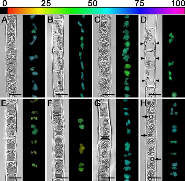
Figure 9.
NIR and corresponding Y(II) images (false colour) of Zygogonium AUT filaments with different cell morphologies. The relative Y(II) as a percentages is indicated by the colour bar at the top. (A) Zygogonium AUT-p. (B) Zygogonium AUT-g. (C) Purple filament with older cells. (D) Cells depositing cell wall material close to cross cell walls (arrowheads). (E) Filament with numerous aplanospores close to cross cell walls of vegetative cells. (F) Encysted cells. (G) Germinated aplanospores as indicated by dark remnant of cytoplasmic residue. (H) Purple filament with one spherical inclusion per cell. Bars = 20 μm.

