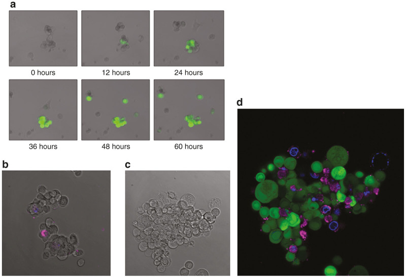
Figure 4.
Fluorescence microscopy demonstrates virus-induced green fluorescent protein (GFP) expression and presumably noninfected bystander cell apoptosis. (a) Images acquired by fluorescent and differential interference contrast confocal microscopy show a tumorsphere on post-infection day 3 following infection with an MOI of 1.0. Cells expressing GFP (green) are infected with NV1066. (b) A merged image of control, uninfected cells expressing only small amounts of Annexin V and DAPI. (c) The control DIC image of the stained tumorsphere. (d) Cy-5 tagged Annexin V-positive cells appear magenta, which indicates phosphatidylserine residues of preapoptotic cells. Cells stained blue have taken up DAPI, indicating increased cellular permeability and a later stage of apoptosis.

