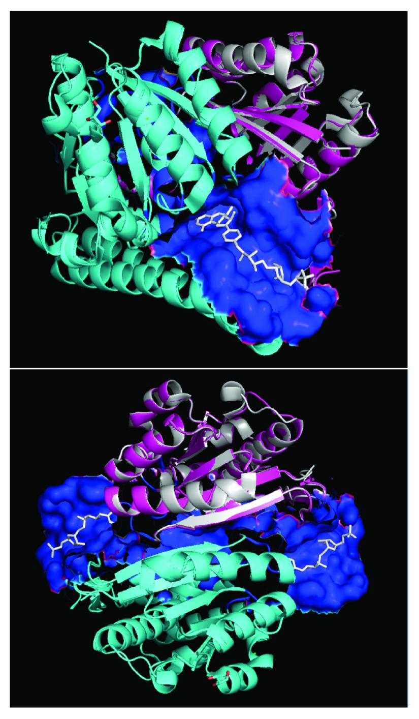
Figure 1.
Modeled quaternary structure of A5UMI1/3IQZB (cyan) and A5UMI1/3IQZF (pink) after respective alignments onto chain-B and chain-F of 3IQZ within PyMOL 28. 3IQZ’s chain-F is highlighted in silver. Dual chain model site residues (blue surface) were inferred from residues in chain-B and chain-F models that are within 7 Å of the 3IQZ ligand (H4M - white). 3IQZ’s chain-B and chain-F form a quaternary structure with two different H4M binding sites (bottom).

