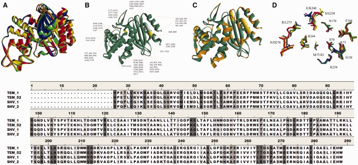
Fig. 1.
(A) Overlay of the ribbon structure of TEM-1 (protein database (PDB) ID: 1ZG4, yellow), TEM-52 (PDB ID: 1M40, red), SHV-1 (PDB ID: 1SHV, green), and SHV-2 (PDB ID: 1N9B, blue). (B) Ribbon diagram of SHV-1 (PDB ID: 1SHV) with amino acid differences with TEM-1 identified, S70 is in yellow. (C) Ribbon diagram of SHV-1 (PDB ID: 1SHV) with the location of amino acid differences with TEM-1 marked in orange. (D) Active site overlay of TEM-1 (PDB ID: 1ZG4, yellow), TEM-52 (PDB ID: 1M40, red), SHV-1 (PDB ID: 1SHV, green), and SHV-2 (PDB ID: 1N9B, blue). The amino acids studied in this article are also shown. Overall, the active site amino acids are very similar between these proteins. The amino acid alignment of these four β-lactamases is also shown below with shading at the site of amino acid differences illustrated in B and C.

