
FIG. 3.
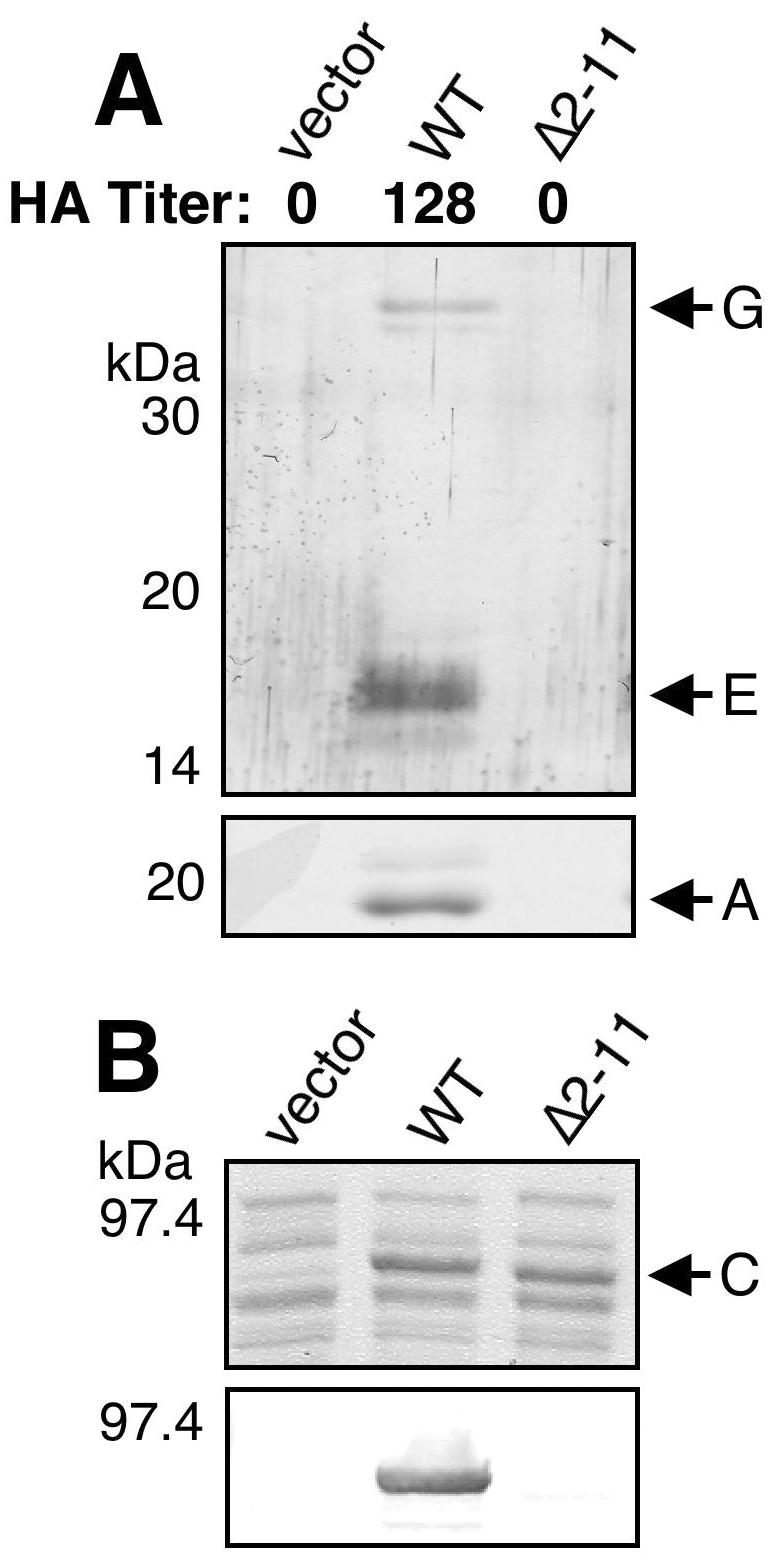
(A) Analysis of pilus biogenesis by PapCΔ2-11. Strain AAEC185/pMJ2 (ΔpapC pap operon) was complemented with the vector alone, WT PapC, or PapCΔ2-11, and assembly of adhesive pili was measured by agglutination of human erythrocytes. The HA titer is the highest fold dilution of bacteria that still provides agglutination. Assembly of pili on the bacterial surface was determined by heat extraction and magnesium precipitation, followed by SDS-PAGE. The rod subunit PapA was detected by Coomassie blue staining (bottom). The tip fibril subunits PapE and PapG were detected by blotting with anti-P pilus tip antibody (top). PapCΔ2-11 was unable to agglutinate erythrocytes or assemble pili. (B) Overlay assay for targeting of PapDG chaperone-adhesin complexes to PapCΔ2-11. OM was isolated from SF100 expressing the vector alone, WT PapC, or PapCΔ2-11. Duplicate samples were separated by SDS-PAGE and either stained with Coomassie blue to show the amount of PapC loaded (top) or transferred to PVDF membrane for the overlay assay (bottom). The PVDF membrane was incubated with PapDG-containing periplasm, and PapDG binding to PapC was detected by blotting with anti-PapDG antibody. The PapCΔ2-11 mutant protein was defective for PapDG binding.