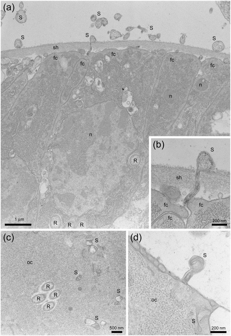
Fig 3. Further images of Spiroplasma and Rickettsia in the female reproductive tissues of M. desjardinsi observed by transmission electron microscopy.
a: Differentiating part of an ovariole, with Spiroplasma-like structure outside and inside the sheath and Rickettsia-like structure in the follicle cells. b: Magnified image of (a), showing the presence of Spiroplasma-like structure in the intercellular space and the sheath, possibly at the timing of intrusion into a follicle cell. c: Cytoplasm of an oocyte, showing Rickettsia and Spiroplasma-like structure. d: Spiroplasm-like structure possibly at the timing of intrusion into the oocyte. S: Spiroplasma; R: Rickettsia; oc: oocyte; fc: follicle cell; sh: sheath; n: nucleus.

