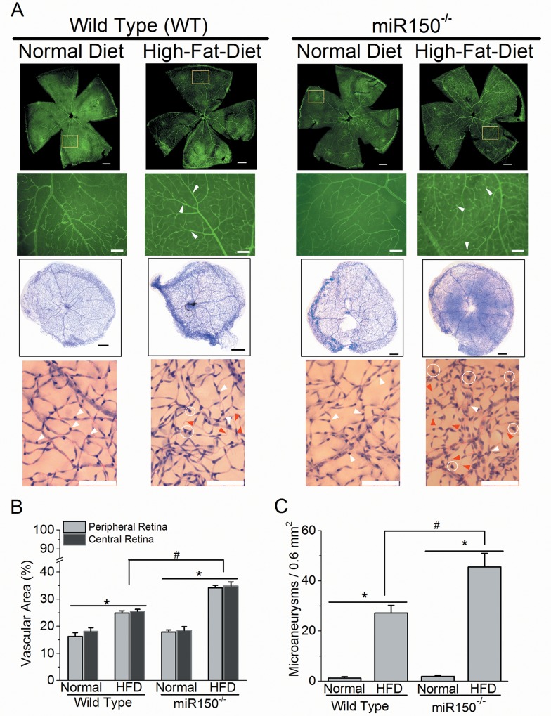
Fig 3. Deletion of miR-150 exacerbates HFD-induced DR neovascularization and microaneurysms.
(A) Upper two rows: the whole mount retinal vasculature was stained with FITC-labeled isolectin-B4. The first row: the fluorescent images from 4 experimental groups were taken at 5X (scale bar = 400 μm). The highlighted regions (yellow square) were magnified at 10X and displayed in the second row. The second row: the fluorescent images from 4 experimental groups were taken at 10X (scale bar = 100 μm). Lower two rows: the mouse retinas were trypsin-digested and the retinal vasculatures were stained with hematoxylin and eosin. The third row: the whole retinal vasculature images are shown (scale bar = 400 μm). The fourth row: magnified images of retinal vasculature (from the third row) are shown (scale bar = 100 μm). White arrowheads indicate the pericytes, the red arrowheads indicate the acellular capillaries, and the white circles indicate the microaneurysm-like (vascular extrusion) structures. (B) Wild type and miR-150-/- mice fed with HFD have significantly higher (*) vasculature in central and peripheral retinal areas compared to mice fed with normal chow diet (Normal). There is a statistical significant difference in the interaction between miR-150 null mutation and HFD regimen (2-way ANOVA; #). (C) Wild type and miR-150-/- mice fed with HFD have significantly higher (*) densities of microaneurysm-like structures (microaneurysms per 0.6 mm2 retinal area) compared to mice fed with normal chow diet (Normal). There is a statistical significant difference in the interaction between miR-150 null mutation and HFD regimen (2-way ANOVA; #). WT-Normal: n = 6; WT-HFD: n = 8; miR-150-/--Normal: n = 6; miR-150-/--HFD: n = 8. p < 0.05 (denoted as *, #).

