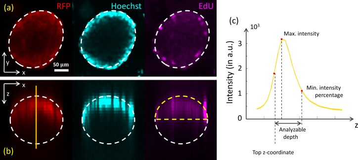
Fig 4. Light attenuation in multi-cellular spheroids.
(a) Top view from the xy-plane of a spheroid with an ellipsoid fitted, where the RFP (561 nm), Hoechst (405 nm), and EdU (640 nm) channels are shown. (b) The same spheroid, in a side view (xz-plane). No signal is detected from the lower part of the spheroid (assuming that the spheroid is of an ellipsoidal shape). (c) Parameters of the spheroid derived from the vertical profile curve of the RFP signal through the ellipsoid center: top z-coordinate, maximum intensity, analyzable depth (corresponding to the user-defined minimum intensity percentage).

